Scoliosis under 45º is mild, but some children may develop spine deformities that continue to get more severe as they grow. This is because of the increase in the angle of deformity. Severe scoliosis can be disabling. An especially severe spinal curve more than 80º can reduce the amount of space within the chest, making it difficult for the lungs to function properly.
Scoliosis cannot be completely cured, but we can freeze the progression of the disorder through braces at early stages (25º to 45º). Surgery is effective in a serious stage.
The main factor of treatment is to build confidence in the person with Scoliosis as they can live a normal life like others. New research results guarantee painless non-invasive procedures (Please refer treatment below).
Recent longitudinal studies reveal that the most common form of the condition, late-onset idiopathic scoliosis, is physiologically harmless and self-limiting even without treatment. The rarer forms of scoliosis pose risks of complications.
Diagnosis of Scoliosis
Early detection of scoliosis is important to freeze the progression of the condition. Recent advancements in research helped scientists to develop effective technologies to detect and treat Scoliosis with less or no complications. This article composes of current methods and procedures involved in the diagnosis of scoliosis.
Currently there are two screening methods to diagnose scoliosis, they are:
- Physical examination
- Imaging studies
Physical examination
Physical examination is done to screen the patient for any abnormality or alteration in normal body structure. It consists of two parts:
-
- Observation
- Palpitation
Observation
In this procedure the examiner will observe the clients body for any visible abnormalities in the body especially in the trunk, ribs, spine, shoulder and waist for asymmetry or abnormal bulging. The widely used physical screening examination is Adam’s Forward Bend Test.
Palpitation
In this procedure the examiner palpates the spine or back bone for any deformation, in case there is no obviously visible alteration, but slight bulging or swelling is present.
Physical Screening examinations
Adam’s Forward Bend Test
In this procedure, the client is asked to bend forward dangling the arms, with the feet together and knees straight. The curve of structural scoliosis is more apparent when bending over. In a client with scoliosis, the examiner may observe an imbalanced rib cage, with one side being higher than the other, or other deformities as shown in the figure (1).

Adam’s forward bend test
Drawbacks of Adam’s Forward Bend Test
Adam’s Forward Bend Test is not sensitive to abnormalities of lowerback, a very common site for scoliosis. As 15% of scoliosis cases are arising from this missed site (lumbar region), many experts do not recommend it as the sole method for screening for scoliosis, especially in the case of lumbar scoliosis.
Other Physical Tests.
Gait examination
The client is assessed for his/her walking style for structural abnormalities of waist, shoulders and extremities.
Musculo-skeletal examinations
The doctor will check leg length and look for tight tendons in the back of the leg, which may cause an uneven leg length or other back problems.
Neurological examination
The doctor will also check for neurological impairment by testing reflexes, nerve sensation, and muscle function.
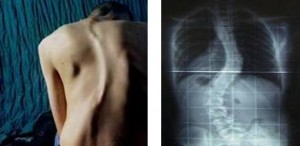
Imaging Tests
X-ray is considered as the most reliable and cost effective way to determine the progress and extent of Scoliosis. T MRI (magnetic resonance imaging) can identify spinal cord and brain stem abnormalities and is useful before surgery for detecting defects that could lead to potential complications.

X-Ray picture of scoliosis
Benefits of X-Rays
If screening indicates scoliosis, the child may be sent to a specialist who takes an initial x-ray and monitors the child every few months using repeated x-rays. X-rays are essential for an accurate diagnosis of scoliosis:
- They reveal the degree and severity of scoliosis.
- They show other spinal abnormalities, including Kyphosis (hunchback) and hyperlordosis (swayback).
- X-rays help the doctor determine whether skeletal growth has reached maturity.
- X-rays taken when patients are bending forward can also help differentiate between structural and nonstructural scoliosis. Structural curves persist when a person bends over, and nonstructural curves tend to disappear. (Muscle spasms or spinal growths may sometimes cause nonstructural scoliosis that shows a curve on bending.)
- Children and young adolescents who have mild curves, and older adolescents, who have more severe curvatures but whose growth has stopped or slowed, need x-rays every few months to detect increasing severity. Young people who are diagnosed with scoliosis should keep their x-rays indefinitely in case they develop back problems later in adulthood and need to be re-examined.
Drawbacks
X-Ray imaging techniques are accurate for detecting scoliosis in the thoracic region, but not in the lumbar region.
Magnetic Resonance Imaging
The benefit of Magnetic resonance imaging (MRI) is that it does not use radiation, as x-rays do. But it is expensive. MRI can identify spinal cord and brain stem abnormalities and is useful before surgery for detecting defects that could lead to potential complications.
Scoliosis detection
The severity of scoliosis is usually determined by two factors:
- The extent of the spinal curvature.
- The Angle of the trunk rotation (ATR)Both are measured in degrees. Scoliosis is diagnosed when the clients (patient) spinal curvature measures 10° or more. If the spinal curve is above 20° the condition requires medical attention. The same applies of the ATR measurement greater than 5°.
| Curve | Treatment |
| 0-20° | Observe for progression |
| 20-25° | Brace if progression documented, and substantial growth remaining |
| 25-30° | Brace if progressive and growth remains |
| 30-40° | Brace if growth remains |
| 40-45° | Brace if growth remains versus surgery |
| >50° | Surgery |
The relation of Spinal curvature and Angle of trunk rotation (ATR)
Spinal curvature: Curvature of the spine is mainly measured by Cobb angle. The Cobb angle is a measure of the curvature of the spine in degrees which helps the doctor to determine what type of treatment is necessary. A Cobb angle of 10 degree is regarded as a minimum angulation to define Scoliosis.
Angle of Trunk rotation: Scoliometer is a device which measures angle of rotation. Results of the Scoliometer can indicate problems, and some experts believe it is a useful device for widespread screening. Scoliometer, however, measure rib cage distortions in more than half of children who turn out to have very minor or no sideways curves. Scoliometer are not accurate enough to guide treatment, and, if results show a deformity, x-rays need to be performed.
Client with a spinal curve of 20° usually have a trunk rotation (ATR) of 5°.
Scoliosis Treatment
Advancement of technology has brought about a lot of changes in the treatment of Scoliosis. Scoliosis is not a disease and the affected person can lead a normal life like others. Clinical treatment varies with the character of Scoliosis. Most of the treatment is based on the inhibition of Scoliosis progression and surgical correction of the deformity.
The clinical management of Scoliosis composes of four parts:
a. Observation
b. Physiotherapy
c. Bracing
d. Surgery
Clinical Management
Observation and mild physical therapy are for the clients who have a moderate size curves from 0 to 20. Many times Scoliosis below 20 is self limiting and doesn’t need medical treatment. The physician will measure the curve on a regular schedule and base treatment decisions on the rate of curvature progression. This is mainly for the clients at growing stage. (Infantile scoliosis, Juvenile Scoliosis and Adolescent scoliosis) .
Physiotherapy
The Schroth method is the widely implemented physiotherapy method for treating scoliosis. It has been used successfully in Europe since 1920.
Schroth method is based on the thesis that scoliosis always involves asymmetrical muscle groups in the back and elsewhere, which in normal bodies are more evenly symmetrical, that can be at least partially corrected by targeted exercises.
It is a noninvasive, physiotherapeutic treatment, developed in Germany by scoliosis sufferer Katharina Schroth. Now as the part of health education, this procedure is taught to scoliosis patients in clinics specifically devoted to Schroth therapy in Germany, Spain, England and North America.
The indications for treatment depend on degree of curvature, maturity of the patient, and the individual curve pattern. While evidence supporting such conservative, non-invasive treatments is weak, today conservative management of scoliosis can be regarded as being evidence-based; no substantial evidence has been found to support surgical intervention.
Bracing
Braces are used to restrict the progress of scoliosis. It restricts the increase in spinal curvature and protects the client from developing complications. There are different types of scoliosis braces:
1. Thoraco-Lumbo-Sacral-Orthosis (TLSO)
In a TLSO brace the posterior portion of the brace extends from the sacrococcygeal junction to just inferior of the scapular spine. This excludes elastic or equal shoulder straps or other strapping. The anterior portion extends from the symphysis pubis to the xiphoid. Some TLSO’s may require the anterior portion to extend up to the sternal notch.
The most common form of a TLSO brace is called the “Boston brace”, and it may be referred to as an “underarm” brace. This brace is fitted to the client’s body and custom molded from plastic. It works by applying three-point pressure to the curvature to prevent its progression. It can be worn under clothing and is typically not noticeable. The TLSO brace is usually worn 23 hours a day and it can be taken off to swim, play sports or participate in gym class during the day. This type of brace is usually prescribed for curves in the Lumbar or Thoraco-lumbar part of the spine.
2. Cervico-Thoraco-Lumbo-Sacral-Orthosis (known as a Milwaukee brace)
The CTLSO brace was originally designed by Blount and Schmidt in 1946 for postoperative care when surgery required long periods of immobilization. The Milwaukee brace (CTLSO) is similar to the TLSO, but includes a neck ring held in place by vertical bars attached to the body of the brace. It is usually worn 23 hours a day. This brace is normally used with growing adolescents to hold a 25° to 40° advancing curve. The brace is intended to minimize the progression to an acceptable level, not to correct the curvature. For corrective measures, special exercises or physical activities are used. If the curvature continues despite the brace, surgery may be required. Like TLSO brace, Milwaukee brace can also be taken off while doing activities during the day. This type of brace is often prescribed for curves in the Thoracic spine.
3. Charleston Bending Brace
Charleston bending brace is otherwise known as “night-time” brace because it is only worn while sleeping. A Charleston back brace is molded to the patient while they are bent to the side. When the child bend against the curve, pressure is generated and it helps to improve the corrective action of the brace. Nighttime only wear frees the adolescent of the negative body image so often associated with this type of treatment, allowing full participation in normal, social, athletic, and academic activities. Curves must be in-between 20 – 40 degree range and the apex portion of the curve needs to be below the level of the shoulder blade for the Charleston brace to be effective.
SURGICAL MANAGEMENT
Surgery is the most effective treatment at present through which the progression of scoliosis is effectively blocked and the condition can be reversed in most cases. Surgical management is the only option for the clients having progressive scoliosis over 50 degree curvature of spine in order to protect internal organs. Surgery is also performed if the deformity is cosmetically unacceptable to the client.
Surgery is the preferred option if,
1. The client is having progressive scoliosis over 50 degree curvature of spine.
2. Deformity is cosmetically unacceptable to the client.
3. Clients with Spina bifida and cerebral palsy
4. Curves that affect physiological functions such as breathing and cardiac functions.

Fig 1.Scoliosis treatment: before and after surgery
The main types of surgeries are:
§ Anterior fusion: In this procedure an incision is made at the side of the chest wall for surgical approach.
§ Posterior fusion: It involves the use of a metal instrument. In this procedure an incision is made on the back for the surgical approach.
One or both of these surgical procedures may be needed. The surgery may be done in one or two stages and, on average, will take four to eight hours.
Spinal fusion with instrumentation
Spinal fusion composes of two parts:
· Fusing the vertebrae along the curve
· Supporting these fused bones with instrumentation (steel rods, hooks, and other devices) attached to the spine

Fig 2.Implimentation of instruments
In this surgical procedure, bone obtained from elsewhere in the body (autograft) or from a donor (allograft) is grafted to the vertebrae. Gradually, they will form one solid bone mass and the vertebral column becomes rigid. This can be performed by anterior (front) aspect of the spine by entering the thoracic or abdominal cavity or, more commonly, performed by posterior (back) aspect. A combination of both aspects may be used in more complicated condition.
Cotrel-Dubousset instrumentation
The technique of using a combination of rods, screws, hooks, and wires fixing the spine, to apply stronger, safer forces to the spine is known as the Cotrel-Dubousset instrumentation. This is the most effective technique available currently.
Thoracoplasty
Thoracoplasty is the procedure of shortening selected ribs in the thoracic or chest area. This procedure is done to reduce the size and severity of a rib hump which may accompany scoliosis. This procedure is also called costoplasty rib hump is evidence that there is some rotational deformity to the spine. Thoracoplasty may also be performed to obtain bone grafts from the ribs instead of the pelvis, regardless of whether a rib hump is present. Thoracoplasty can be performed as part of a spinal fusion or as a separate surgery.
Surgery without fusion
Researches and studies conducted on Scoliosis are novel and promising. Advanced proimplants that aim to delay spinal fusion and to allow more spinal growth in young children have been developed. The cases in which thoracic insufficiency compromises physical ability to breathe and applies significant cardiac pressure, ribcage implants that push the ribs apart on the concave side of the curve may be especially useful. These vertical expandable prosthetic titanium ribs (VEPTR) provide the benefit of expanding the thoracic cavity and straightening the spine in all three dimensions while allowing it to grow.
********************************************************************************************
References:
Scoliosis: Causes, Tests and Treatments by John Hewitt MA and Mohamed Awad MD
Scoliosis and the Human Spine – National Scoliosis Foundation
The National Center for Biotechnology Information – U S A
