The Cell Cytoskeleton: Deposing the Blob Myth
Cells, the most basic units of life we each studied in school, are complex machines dumbed-down to be membrane-enclosed, fluid-filled blobs. The beautiful compartmentalization of the eukaryotic cell is unseen by the average Joe. Hundreds of metabolic reactions each mediated by a specific enzyme carried out within a membrane-enclosed space occur each second within the cell to keep said Joe alive in his state of blissful ignorance. This being said, would the idea that the complexity of the cell runs even deeper bring Joe out of his stupor? No – probably not. Yet, within the cell, the cytoskeleton creates an orderly, but dynamic structural framework that makes the cell far from blob-like.
Structure and Composition
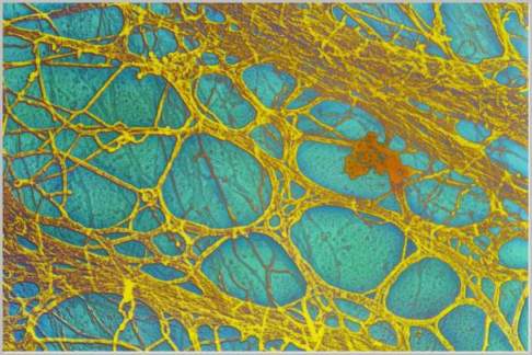
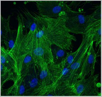
The cytoskeleton of most eukaryotic cells is composed of three basic structural components collectively called cytoskeletal elements: microtubules, intermediate filaments, and microfilaments. The cytoskeletal elements are composed of different proteins; differ in size, and each carries out specific functions within the cell. These elements run throughout the cell cytoplasm creating a highly organized yet changeable structure.
Microtubule
The largest of the cytoskeletal elements is the microtubule. As the name implies, microtubules are protein tubes composed of globular subunits called tubulin that create a helical tube-like structure. These monomers are held together by weak, noncovalent protein-to-protein interactions such as hydrogen bonds or van der Walls interactions. These weak associations allow for rapid disassembly and reassembly in new locations based on the changing needs of the cell. Examples of structures within the cell composed of microtubules include the mitotic spindle, centrioles in animal cells, cilia and flagella, and basal bodies. Microtubules function to maintain cell shape through compression resistance and are important in several aspects of cell motility.
The mitotic spindle forms during the changes that occur in the cell cycle. Kinetochore microtubules of the spindle attach to the chromosomal centromeres allowing the chromosomes to be moved to the metaphase plate of the cell. During anaphase these microtubules separate the sister chromatids ensuring that each new daughter cell receives its full genetic compliment of chromosomes. The nonkinetochore microtubules of the spindle elongate the cell preparing it for cytokinesis.
Cilia and flagella are composed on nine doublets of microtubules arranged in a ring surrounding a central pair of microtubules. This is known as a “9 + 2” structure. These doublets are attached to one another with accessory proteins. Specialized motor proteins called dynein arms allow for the locomotive action of cilia and flagellum. Upon phosphorylation through the breakdown of ATP, adenosine triphosphate, the dynein arms undergo conformational shape changes that cause the doublets of microtubules to slide past one another bending the cilia or flagella.
Composed of microtubule triplets are centrioles and basal bodies. This structural arrangement includes nine triplets in a ring held together by accessory proteins. The centrioles in animal cells are involved in the formation of the mitotic spindle. Basal bodies anchor the cilia and flagella within the plasma membrane.
Another important function of microtubules involves the movement of organelles within the cell cytoplasm. The organelles move along microtubules with the aid of specialized motor proteins. These motor proteins associate with certain organelles such as vesicles in a receptor-mediated fashion. Energized by ATP, the motor protein then “walks” the organelle to its new destination within the cell cytoplasm. Important to this process is the changing nature of the cytoskeleton. New microtubules can be quickly assembled to move substances to far-reaching sites within the cell.
Intermediate Filaments
The next cytoskeletal elements according to size are the intermediate filaments. These filaments, found in some eukaryotes such as the vertebrates, provide tension resistance in the structural framework of the cell and are much more permanent features of the cytoskeleton. These cytoskeletal elements exhibit the most structural diversity since they are composed of different proteins in differing cell types. The fibrous subunits that compose intermediate filaments in epithelial cells are several types of keratin. These subunits form stronger associations, an example being disulfide bridges, than those seen in microtubules or microfilaments. As a result, these elements are much stronger and serve the primary role of maintenance of cell shape. They also compose the nuclear lamina, a structural framework within the eukaryotic nuclear envelope, which provides protection for the cell’s genetic material and may function to organize it. Another cell type that contains intermediate filaments is the neuron. These cells’ intermediate filaments are called neurofilaments and are composed of several protein subunits. The elongate axons of neurons are maintained by these cytoskeletal elements. Muscle cells also contain intermediate filaments but these are composed of desmin subunits. Regardless of cell type, the intermediate filaments function to maintain cell shape.
Microfilaments
Microfilaments, the smallest cytoskeletal elements, are primarily responsible for changes in cell shape. These elements are composed of globular subunits called actin that form weak associations as described previously. Two actin chains twist around one another to form a helical, threadlike structure recognized as a microfilament. Actin microfilaments are often associated with motor proteins called myosin as seen in muscle cells. Muscle contraction occurs as actin microfilaments interact with myosin motor proteins that have been energized by ATP.
Microfilaments form a framework directly below the plasma membrane giving the outer portion of the cytoplasm a gel-like consistency while the inner portion of the cytoplasm is more fluid. These regions of the cytoplasm are known as the gel and sol regions, respectively. Two types of cell motility utilize the gel-sol nature of the cytoplasm: amoeboid movement and cytoplasmic streaming. Amoeboid movement, seen in some protozoa and some animal cell types, involves the formation of extensions of the plasma membrane called pseudopodia. These extensions have weakened regions of the gel-type of actin microfilaments. Through interactions with myosin, cell contractions force sol-type cytoplasm into these extensions. In plant cells, similar gel-sol interactions allow for the movement of the cytoplasm that may aid in the distribution of materials throughout the cell. Another important function of microfilaments is the formation of the cleavage furrows seen during animal cell division. During the process of cytokinesis, a contractile ring of actin forms, which functions to pinch the cell into two daughter cells.
Whether maintaining the cell’s shape, aiding in the transport of materials within the cell, or providing cell motility, the cytoskeleton is a dynamic and necessary structure for the eukaryotic cell. Far from being a shapeless blob, the cell is a highly organized structure capable of diverse functions. Adding to this functional diversity is the cytoskeleton, the often-overlooked framework. So Joe what do you think now?
References:
Campbell, Neil and Reece, Jane. Biology, 7th Edition. Benjamin Cummings, San Francisco, 2005.
Alberts, et al. Molecular Biology of the Cell, 4th Edition. Garland Science, New York, 2002.
